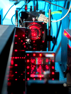
Microscopic move could bring new Nobel knocking
 The researcher awarded the Nobel Prize for changing the world of microscopy may have done it again.
In October this year, Eric Betzig shared the Nobel Prize in chemistry for his ground-breaking work on high-resolution microscopes – handed one of the highest honours for something designed and built on a friend's living room floor.
But now, Dr Betzig has reported on a new microscopy technique, developed in much more sophisticated surrounds.
The device developed by a team at the Howard Hughes Medical Institute's Janelia Research Campus allows them to observe living cellular processes at world-beating resolution and speed.
Dr Betzig said he came up with his Nobel-winning PALM microscope after he grew frustrated with the limitations of other microscope technologies.
Similarly, he says the new ‘lattice light-sheet microscopy’ was the result of his eventual boredom with PALM.
“Again, I just started to understand the limits of the technology,” Betzig said.
PALM allowed new insight into living systems, but was only effective when they were barely moving, and could not take measurements quickly enough to get high-resolution pictures of fast processes such as cellular division.
Dr Betzig said trying to monitor in real-time with PALM was like trying to watch a football game presented in a few still frames.
“You can see images of a pass, and a touchdown, and of the cheerleaders doing a pyramid. But the rules of the game would only become clear once you saw a game on video,” he said.
“I'd been looking at those pictures my whole life... it was time to take a look at the living stuff in action.”
The new microscope generates a sheet of light coming in from the sides of the sample. The series of beams allows a more rounded view and does less harm to the sample than one solid beam of light.
Researchers using the ‘lattice light-sheet’ technique can snap a high-resolution image of an entire section of a sample, rather than the clear focal point but plurry edges offered by PALM.
The research team has managed to film and present videos of 20 different biological processes in never-before-seen clarity, just a tiny sampling of what the technique will accomplish.
Their paper has been published in the AAAS journal Science.


 Print
Print